Cell Death and Apoptosis
Overview
Cells that do not receive the proper supplies will die, through a step wise process called apoptosis. This is a normal physiologic phenomenon and in fact apoptosis is crucial to the normal development of the nervous system. But halting apoptosis when it is producing degenerative change in the nervous system is now a prime goal for researchers trying to design effective treatments for ALS as well as for other neurological disorders.
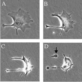
Time lapse photography of a cell undergoing apoptosis. Guy Whitley, Reproductive & Cardiovascular Disease Research Group, St. George's Hospital Medical School, London, U.K.
What is Apoptosis?
Programmed cell death is a series of events where a cell essentially shuts itself down and is eliminated. It is normal for many cells to die of apoptosis as the nervous system forms; it is part of constructing appropriate connections. Apoptosis is crucial for normal brain development and function, as researchers have discovered over the past decade or two. Just as a fruitful orchard results from judicious pruning, effective and efficient connections within the nervous system result from apoptosis.
Apoptosis and ALS
Apoptosis is a sequential process of shutting down a cell that is no longer needed or no longer working properly. By contrast to apoptosis, necrosis results from a direct injury to the nervous system, or acute infection. Necrosis produces an explosion of cell contents accompanied by inflammation and immune activation. Unlike the messy death of infected or traumatized cells in necrosis, apoptosis is a process of carefully choreographed steps to self destruction.
Apoptosis calls into play a cascade of intracellular events to decommission the cell. Scientists now believe apoptosis may play a role in the disease process of ALS, and also in many other neurodegenerative conditions such as Huntington's disease, and is even implicated in the spreading damage after a stroke. Something in the initial disease process in these diverse conditions is triggering apoptosis. Stopping it may be a key factor in designing effective therapy for ALS and many other neurological conditions.
What Triggers Apoptosis?
Stress such as radiation or chemical exposure can trigger apoptosis, scientists have determined, as can infection, or oxidative damage. When certain proteins dock at the cell surface, they are messengers that something in the cell's environment is amiss, and the messages induce apoptosis. Stress produces an increase in certain proteins inside the cell that also initiate apoptosis.
Apoptosis avoids unwanted cell proliferation, which could turn into cancer, or spread a viral infection. Thus, signals that say the cell is not acting properly are natural responses to produce apoptosis, to remove the abnormal cell before it can spread damage.
Cascade of Cellular Events
In apoptosis, a cascade of events comes into play to remove a cell. In early stages, a set of enzymes called caspases begin to work. These proteins break apart the scaffold within the cell. In turn, caspases activate enzymes that take apart DNA.
The cell at this point is visibly undergoing self destruction, as can be viewed through a microscope. Its shape changes, matching the biochemical changes taking place inside. As the cell skeleton is disassembled, the cytoplasm shrinks. The nucleus also shrivels.
Various characteristics of apoptosis show that this process is underway. These apoptotic markers allow investigators to see clearly that apoptosis is the cause of cell death. Aside from the biochemical cascade within the cell, visible signs of an apoptotic cell include broken off bits of the membrane, and small vesicles called apoptotic bodies.
Changes at the cell surface of an apoptotic cell meanwhile signal the macrophages to come and engulf it. This clean removal of the cell avoids the inflammatory problems of necrosis.
Death Receptors
Docking sites on the surface of a cell receive signals that can tip the balance of inner chemistry towards apoptosis. One signal is carried by tumor necrosis factor alpha (TNFa). This signal is generated by immune system cells such as T cells and activated macrophages. TNFa produces further immune activation, and it also can play into apoptosis, though it is not a particularly strong inducer of cell death.
Certain drugs are known to interfere with TNFa, including thalidomide. Drugs called histone deacetylase (HDAC) inhibitors are other promising candidates that affect apoptosis. Some of these drugs have been or will be tested in clinical trials for ALS, to see if they might help delay the disease process.
Mitochondria and Apoptosis
The cell organelles (little organs) called mitochondria (see also mitochondria) are the umpires of apoptosis, they can call the play into action or delay it. The mitochondria contain enzymes that produce apoptosis, and ones that stall the process. For instance, the Bcl-2 family of proteins put off apoptosis. These enzymes are located in the outer membrane of mitochondria.
Release of cytochrome C from mitochondria can activate caspases and produce cell death. Mitochondria show damage early in the ALS disease process, a finding that is leading researchers to study these cell components intensively. Important clues on the roles of mitochondria and apoptosis in ALS undoubtedly will surface as scientists probe further into the details of programmed cell death during the disease.
Recursos en español disponibles. Por favor llame al (212) 619-1400. Gracias.
DONATE